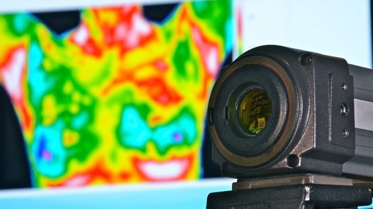
Red and White indicating significant abnormal blood flow and risk
As with any technology, as time progresses better thermography cameras and software is developed increasing the detection ability and resolution thus making evaluating the images more precise. As early as 1976, at the Third International Symposium on Detection and Prevention of Cancer in New York, thermography was established by consensus as the highest risk indicator for the possibility of the presence of an undetected breast cancer. It also has been shown to predict subsequent occurrences [1-3]. The Wisconsin Breast Cancer Detection Foundation presented a summary of its findings in this area, which has remained undisputed [4]. This, combined with other reports, has undeniably vouched that thermography is the highest risk indicator for the future development of breast cancer and is 10 times effective where there is a family history of the disease [5]. This additional effectiveness is in great part due to patients being aware of their predisposition and proactively obtaining screening. Thermography is an ideal choice with no pain and no radiation in where frequent screening by other methods could result in radiation overexposure.
A 10,000 women study by Gautherie found that, when applied to asymptomatic women, thermography was very useful in assessing the risk of cancer by dividing patients into low- and high-risk categories. This was based on an objective evaluation of each patient’s thermograms using an improved reading protocol that incorporated 20 thermopathological factors [6].

Thermography Screening indicating the possibility of Fibrocystic Breast
Therograph Screening indicating the possibility of Fibrocystic BreastFrom a patient base of 58,000 women screened with thermography, Gros and associates followed 1,527 patients with initially healthy breasts and abnormal thermograms for 12 years. Of this group, 40% developed malignancies within 5 years. The study concluded that “an abnormal thermogram is the single most important marker of high risk for the future development of breast cancer” [7].
Spitalier and associates followed 1,416 patients with isolated abnormal breast thermograms. It was found that a persistently abnormal thermogram, as an isolated phenomenon, is associated with an actuarial breast cancer risk of 26% at 5 years. Within this study, 165 patients with non-palpable cancers were observed. In 53% of these patients, thermography was the only test which was positive at the time of initial evaluation. It was concluded that: (1) A persistently abnormal thermogram, even in the absence of any other sign of malignancy, is associated with a high risk of developing cancer, (2) This isolated abnormal also carries with it a high risk of developing interval cancer, and as such the patient should be examined more frequently than the customary 12 months, (3) Most patients diagnosed as having minimal breast cancer have abnormal thermograms as the first warning sign [8-9].
References
[1] Amalric, R., Gautherie, M., Hobbins, W., Stark, A.: The Future of Women with an Isolated Abnormal Infrared Thermogram. La Nouvelle Presse Med 10(38):3153-3159, 1981
[2] Gautherie, M., Gros, C.: Contribution of Infrared Thermography to Early Diagnosis, Pretherapeutic Prognosis, and Post-irradiation Follow-up of Breast Carcinomas. Laboratory of Electroradiology, Faculty of Medicine, Louis Pasteur University, Strasbourg, France, 1976
[3] Hobbins, W.: Significance of an “Isolated” Abnormal Thermogram. La Nouvelle Presse Medicale 10(38):3153-3155, 1981
[4] Hobbins, W.: Thermography, Highest Risk Marker in Breast Cancer. Proceedings of the Gynecological Society for the Study of Breast Disease. pp. 267-282, 1977.
[5] Louis, K., Walter, J., Gautherie, M.: Long-Term Assessment of Breast Cancer Risk by Thermal Imaging. Biomedical Thermology. Alan R. Liss Inc. pp.279-301, 1982.
[6] Gauthrie, M.: Improved System for the Objective Evaluation of Breast Thermograms. Biomedical Thermology; Alan R. Liss, Inc., New York, NY; pp.897-905, 1982
[7] Gros, C., Gautherie, M.: Breast Thermography and Cancer Risk Prediction. Cancer 45:51-56, 1980
[8] Amalric, R., Giraud, D., et al: Combined Diagnosis of Small Breast Cancer. Acta Thermographica, 1984.
[9] Spitalier, J., Amalric, D., et al: The Importance of Infrared Thermography in the Early Suspicion and Detection of Minimal Breast Cancer. Thermal Assessment of Breast Health (Proceedings of an International Conference), MTP Press Ltd., pp.173-179, 1983
